Syllabus
Practical Macromolecular 3D Structure Visualization & Structural Bioinformatics
A Two-Morning Workshop -- University of Massachusetts, Amherst

Monday June 13 & Wednesday June 15, 2016.
9:00 AM - 1:00 PM each day, Fine Arts Center 444.


Bring Your Laptop Computer, Please!

Taught by Eric Martz, Ph.D.
principal author of FirstGlance in Jmol ( buttons in the journal Nature) and MolviZ.Org,
team member of Proteopedia.Org, and coauthor of the ConSurf Server.


This document is on-line:
Workshops.MolviZ.Org

Please see the Workshop Announcement

Objectives: Participants will use highly user-friendly software for visual investigation of 3D molecular structures of proteins, nucleic acids, and their interactions with each other and with ligands, substrates, and drugs; and of protein evolutionary conservation. Hands-on experience will be largely with molecules of each participant's choosing. The software easily makes high quality molecular images and rotating molecules for Powerpoint slides.

Software: All software in this course is free, and works on Windows, Mac OS X, or linux. The major tool is FirstGlance.Jmol.Org, adopted by Nature ( buttons) and other journals and resources.

Level & Pace: This workshop is designed for faculty, postdocs, research staff and graduate students familiar with basic biochemistry, but with no previous molecular visualization software experience. It progresses rapidly to powerful tools that will be of interest to specialists in protein structure and bioinformatics. Experienced participants are encouraged to work at their own speed, ahead of the group -- there is plenty of power to discover within these tools!

        Get Started:
            Use the Firefox browser
            Optional: Installing and Enabling Java

    Please get started on your own while the group gets settled:

  1. If you brought your own laptop, you are welcome use it. (iPads will be too slow.)
  2. Use the Firefox browser (Why?). If you do not have it, take a few minutes to install it (firefox.com).
  3. Go to our syllabus: Workshops.MolviZ.Org.
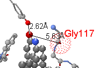
  4. Click on today's syllabus. Now you can see this document in your browser.
  5. Go to Atlas.MolviZ.Org.
  6. In the Atlas, choose any molecule deemed Straightforward and click on the link to FirstGlance. After a minute or so to load, you should see a rotating molecule. Have a look around at the information, views and tools in FirstGlance.
  7. If you have any difficulty or the molecule does not appear, or does not rotate, ask for help!

  8. OPTIONAL: Installing and Enabling Java

    You can see molecules without Java, using JSmol, in the primary tool of this course: FirstGlance.Jmol.Org. For some older sites that display molecules in Jmol, or for especially large molecules, you may need to install and enable Java.

    Java will also make FirstGlance run faster and smoother.

    In this course, there are resources that use all of the 3 forms of Jmol that work in web browsers:
    • JSmol (no Java)
    • Jmol_S (signed Java applet)
    • Jmol (unsigned Java applet, deprecated)
    See four forms of Jmol.

    1. Follow these instructions for installing/updating and enabling Java.
    2. TA's and Instructors will help you!
    3. Can you see this Gal4:DNA complex with Java (Jmol_S)?

    4. If you got Java to work, use the Preferences tab in FirstGlance to make Java the default. Then this link to Gal4:DNA should use Java (Jmol_S).

    5. Some sites use an older, unsigned Jmol Java applet. An example is this explanation of Protein Secondary Structure. Can you see the molecule? Make sure to Enable Unsigned Java Applets for this website. Later in the course, you may want to enable other websites.

  9. Workshop Overview

  10. Workshop Overview (Powerpoint Slides)

  11. Nikhil Malvankar (Physics) took this workshop in 2012. Published his homology model in 2015.

  12. Protein Data Bank & PDB Codes
    Proteopedia.Org
    Crystallographic Resolution

  13. The Protein Data Bank (PDB) -- World Wide: -- USA:RCSB -- Japan:PDBj -- Europe:PDBe
  14. PDB identification code examples:


  15. Proteopedia.Org:
    1. Main page: green links connect text to molecular scenes.
    2. Molecules explained by users. Example:
    3. Explanations of structural biology terms and concepts, e.g. asymmetric unit, Protein Data Bank, hydrogen bonds, temperature value, and more: About Macromolecular Structure.
    4. Pages in Chinese, Japanese, Arabic, Turkish, Russian, etc.

  16. X-Ray Crystallography and Resolution


    Finding molecular models of interest
    Each participant should find a molecule of personal interest.

  17. Finding molecular models of interest:
    Each person: please find a 3D model of a molecule related to your research or interests.

    You will use the model that you select for the rest of the workshop.
    How To Find Models:

    Review of Protein Chemistry and Structure.
    We'll do this quickly. The links are for participants who are educators.
    Standard amino acids.
    Click to see details.

  18. Central Dogma: DNA mRNA Protein.     DNA structure in Jmol / Estructura del ADN
  19. 20 Amino acids
  20. Polypeptide chain geometry and steric restrictions
  21. Covalent and non-covalent chemical bonds
  22. Typical hydrogen bond within a protein: hydrogen donor atom is covalently bonded to hydrogen; acceptor atom is not. In proteins, donor-acceptor distance can be 2.5 to 3.5 Å.
  23. Secondary Structure
  24. Folding: hydrophobic collapse
  25. Protein folds cannot be reliably predicted from sequence alone (using ab initio theory).
  26. Fold does not always equal function: About 10% of proteins are thought to be fully disordered to support their functions, and 40% of eukaryotic proteins have at least one long disordered region. This is termed intrinsic disorder. Examples: 1jsu, 2rrl.

  27. Evolutionary Conservation: ConSurfDB & ConSurf
      Enolase (4enl; a glycolytic enzyme) evolutionary conservation from ConSurf. Catalytic cleft is at the left. More..

  28. Identify Functional Sites In Your Molecule Using The ConSurf Server:
  29. Get your ConSurf calculation started while we discuss evolutionary conservation:

    1. Go to the ConSurf Server.
    2. Select Amino Acids.
    3. Check YES there is a known protein structure.
    4. Enter the PDB code, or upload your homology model.
    5. Select a chain for analysis. ConSurf can analyze only one chain at a time.
    6. Check NO, you will not be uploading a multiple sequence alignment. (ConSurf will create one for you.)
    7. Leave all parameters at their pre-set defaults. (OPTIONAL: If you want a research grade result, check "Let me select the sequences for the analysis manually out of BLAST results", and see Limiting ConSurf Analysis to Proteins of a Single Function.)
    8. Enter your email address at the bottom of the form (this is important so you don't lose your results). Optionally enter a job title.
    9. Click the button Submit. It may take an hour or more to complete this calculation.
    10. When it is finished, under Final Results, click the link View ConSurf Results with FirstGlance in Jmol.
    11. Example: 4enl result in ConSurf.

  30. Background: see Introduction to Evolutionary Conservation.

  31. Effect of mutation on protein function Genetic consequence Example
    Function LOST** CONSERVED:
    mutation LOST from gene pool
    R133C*
    None NOT conserved:
    mutation remains in gene pool
    E143?*
    * in methyl CpG binding protein 2 (MeCP2), 3c2i:

       ASASPKQRRS IIRDRGPMYD DPTLPEGWTR KLKQRKSGRS AGKYDVYLIN
       PQGKAFRSKV ELIMYFEKVG DTSLDPNDFD FTVTGRGSPS RHHHHHH
             ^          ^

    ** R133C causes Rett syndrome, a severe neurological disorder.
    Gray: disordered in crystal, absent in model 3c2i.


    1. Enzyme example: ConSurf-colored sequence -- 4enl ConSurf Result -- enolase in Wikipedia.
    2. Multiple sequence alignments reveal conservation: MSA for 4ENL in black and white (printed handout).
    3. Detail of MSA with color. ConSurf does a much more sophisticated job of calculating evolutionary conservation scores than this simple example!
    4. On the less conserved side of the molecule, touch the isolated highly conserved residues to display the amino acid and sequence number. Gly236 and Pro290 are highly conserved. Why?.

  32. ConSurf's Mechanism: Simplified;   Details;   Technical.
  33. Note the Caveats in Proteopedia's Evolutionary Conservation.
  34. There are two ConSurf Servers:
    1. ConSurfDB (DataBase) NOT WORKING IN JUNE, 2016
      • Pre-calculated for every chain in the PDB.
      • Results are shown in Proteopedia.
      • Multiple Sequence Alignments typically include proteins of more than one function, so some conservation may be hidden.
    2. ConSurf
      • Set up each job by hand.
      • Easily select sequences for a single protein function, revealing conservation (within a family of proteins performing a single function) that may be hidden in ConSurfDB.

  35. If you have a serious interest in the conservation in your protein,
    FirstGlance in Jmol: Easy Visualization of Any Macromolecule

    Terminology: "visualization" vs. "modeling". (Light modeling tools)

  36. To start FirstGlance, google "firstglance" (no space), or go to FirstGlance.Jmol.Org. Then enter the PDB code, or upload your homology model.

    Unusually large models may take a long time to display and be sluggish to manipulate in FirstGlance. For such cases, using Java will enable much better performance in FirstGlance. Java is not needed for most models. Ask the instructor for advice. Here are instructions for Installing and Enabling Java.

  37. Explore 1izh in FirstGlance.
    1. Introduction
    2. Molecule Information Tab
      1. Year, Method.
      2. Resolution.
      3. Free R.
      4. Chain details.
      5. Sequences: Crystallized vs. Full Length. Alignment at UniProt (1d66).
      6. Abstract.
      7. Citations.
      8. Text contents of the PDB file.
    3. Views tab
      1. Top 3 rows of views:
        Secondary Structure / Cartoon / N->C Rainbow
        Composition / Hydrophobic/Polar / Charge..
        Local Uncertainty / Vines / Thin Backbone
      2. Buttons.
        Ligands+ / Water / Slab
      3. 1pgb: Hydrophobic core: Hydrophobic/Polar, then Slab.
      4. 1pgb: Amphipathic helices and strands. (In FirstGlance, use Isolate.. on each end of a helix or strand.)
          Potassium channel (1R3J) showing membrane surface planes (from OPM). See the Resources tab in FirstGlance.
      5. Compare with the Hydrophobic/Polar View of 1bl8 or 7ahl.
    4. Resources tab
      1. See lipid bilayer boundaries (1bl8 or 7ahl).
    5. 1pgb: Tools tab with Views.
      1. Salt bridges.
      2. Cation-pi interactions.
      3. Distances.
      4. Salt bridges in Charge View (Red sidechain (-) touching blue sidechain (+)).
      5. Charges with Slab on.
      6. Sidechain distributions in Vines View (rings buried; charges on surface).
      7. Find (review Chart of AA): PHE, (VAL,LEU,ILE), ASN, THR

  38. Explore 9ins in FirstGlance.
    1. Tools tab
      1. Disulfides/S/Se


    Contacts: Anti-Alzheimer's drug analog (*) interaction with acetylcholinesterase (Dvir et al./Sussman, 2002).
  39. Explore 3onz in FirstGlance. (Letter O not numeral zero!)
    1. Molecule Information Tab
      1. Two chains, not sequence identical.
      2. Missing residues.
      3. Ligands+ and non-standard residues
    2. Views tab
      1. Ligands button; smaller ligands.
      2. Hide (chain, toluene, isolated His).
    3. Tools tab
      1. Contacts
      2. Non-covalent interactions for HEM in chain A (blue chain).
    4. Resources tab
      1. Biological unit.

  40. Solution Nuclear Magnetic Resonance (NMR)

    Introduction to Structural Bioinformatics and Genomics
    Educators: You are welcome to use the slides linked below, or to adapt content from them into your own slides.

  41. Slides Covering:     (from bioinformatics.molviz.org)

We won't have time to go through the following resources in detail, so the links and information below are provided mostly for you to use, if you wish, after the workshop.
    Polyview-3D for Animated Powerpoint Slides and Publication-Quality Images:
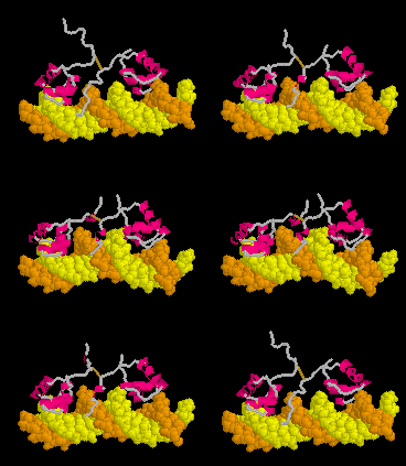
      PowerPoint-Ready Animation from Polyview-3D. Click on the image for a larger view and explanation.

  1. Make Animated PowerPoint Slides and Publication-Quality Images easily with Polyview-3D.
    Proteopedia.Org (Part II): Authoring

  2. Add Molecular Scenes and Content to Proteopedia.Org
  3. Its a wiki: you or your students can add pages or customized molecular scenes in Jmol.
  4. Great for journal supplementary materials or research group websites as well as molecular structure tutorials and student reports.
  5. Protect your pages from being changed by anyone else, e.g. Nucleosomes (protected).
  6. An easy Scene Authoring Tool attaches your customized views to Green links.
  7. This is, by far, the easiest place to create molecular tutorials, e.g. Nucleosomes (publically editable).
  8. Sandboxes (enough for your entire class!) allow students to try authoring temporary molecular scenes -- without individual accounts. Screenshots can document student work. See Teaching Strategies Using Proteopedia.

  9. Jmol in Scientific Journals:

  10. Interactive 3D Complements in Proteopedia: "Supplementary Materials"

  11. FirstGlance in Jmol: Look for the buttons in Nature and Nature Structural and Molecular Biology.

  12. "Jmolized" Interactive 3D Journal Figures:
    Educational Resources

  13. Tutorials disponible en español at MolviZ.Org (Estructura del ADN; Modelo de bicapa lipídica y canal de gramicidina; Estructura del agua: enlaces de hidrógeno) and BioModel (Estructura de proteínas; Glúcidos, Lípidos, Vitaminas, Aminoácidos, Hélice alfa, Hebra beta, Lisozima, Nucleósidos, ADN, ARN, et al.)

  14. HighSchool.MolviZ.Org: Resources for High School Teachers.
  15. Toobers in Science Education!
  16. Molecular Workbench (from Concord Consortium): Activities for High School Students with built-in assessment and reporting.

    If this image is not moving, reload the page!
    Morph of the lactose repressor bending DNA as it recognizes the operon. More..
  17. Teaching Scenes, Tutorials, and Educators' Pages in Proteopedia, including Molecule of the Month.
  18. Teaching Strategies Using Proteopedia.
  19. MolviZ.Org: Martz Central: Resources for High School, College, and Researchers.

  20. Bird Flu: N1 vs. Tamiflu Lesson Plan:
  21. Animations / Morphs: Conformational Changes (see MOVIE at right).

    Molecular Modeling (Time Permitting, or Later Private Sessions)

  22. Mutating your model:
    1. Changing residue sidechains and rotamer minimization with DeepView.
    2. DeepView beginners should start with the superb Molecular Modeling for Beginners by Gale Rhodes, Univ. Southern Maine.

  23. Structural Alignment of two or more chains or molecules, and how to view the alignment.


Keep in touch!